Specialty: Uroradiology
Modalities: Ultrasound, Plain Film X-ray, Nuclear Medicine Imaging, Magnetic Resonance Imaging (MRI), Computed Tomography (CT)
Radiologists: Vijay Gudla, Dr Grainne Murphy, Dr Ben Miller, Dr Arul Ganeshan, Dr Paul Crowe
 Uroradiology is concerned with imaging disorders of the urinary tract for Urologists and Renal Physicians. This includes disorders of the kidneys, ureters, and bladder, in men and women and in also men, the testes, prostate gland and the penis.
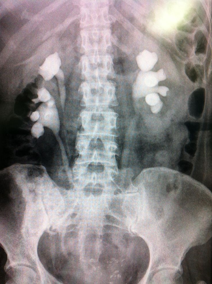
We typically might be asked to image patients with blood in their urine, difficulty passing urine, or pain in the kidney or bladder areas. Kidney stones, infections and cancers of the urinary tract are typical problems encountered.
A range of modern imaging modalities are used to investigate patients with symptoms and signs and are widely used to diagnose these kinds of conditions and to help plan and guide their treatment.
Examples of Examinations performed
Ultrasound Kidneys & Bladder
Often a first step to look for obvious abnormalities
CT KUB
A scan (using X-rays) of the abdomen and pelvis to look for kidney stones
CT Urogram
Like the above but with an injection of X-ray dye to better show the kidneys, ureters and bladder.
MRI Prostate
Uses a magnetic field and radiowaves to obtain images of the prostate gland.
Nuclear Medicine Bone Scan
An injection of a small dose of radioactive dye to look for any evidence of disease in the bones of the skeleton.
Interventional Radiology
Uses ultrasound or X-rays to guide treatments which often previously needed surgery. Examples include obtaining samples of tissue (biopsy) unblocking kidneys (nephrostomy), removing kidney stones (percutaneuos nephrolithotomy – PCNL) and treating suitable kidney tumours with high frequency energy (Radiofrequency Ablation – RFA).
The Urology MDT is held every Tuesday in the radiology Seminar room at 13:00. Here we discuss the scans and any pathology and plan patients treatments.